Quick-Neuron™ Excitatory – Human iPSC-Derived Neurons (Healthy Donor)
Quick-Neuron™ human iPSC-derived excitatory neurons are off-the-shelf glutamatergic neurons that provide a consistent, biologically relevant in vitro model of human excitatory neuronal function. These cryopreserved, ready-to-use iPSC-derived neurons mature rapidly in culture and deliver reproducible performance for studying synaptic activity, neuronal network formation, disease modeling, drug discovery, and neurotoxicity screening.
$850.00
Advantages of iPSC-Derived Excitatory Neurons
Rapid Differentiation
~7 days
Functionally Validated
QC confirmed
Highly Pure Population
VGLUT1+, TUBB3+
No Genetic Footprint
0 modifications
Excitatory Neuron Protocol
Our easy-to-follow, optimized excitatory neuron protocol supports rapid and reproducible differentiation of human induced pluripotent stem cells (iPSCs). This step-by-step guide outlines key culture stages and timelines for generating functional excitatory neurons from human iPSCs.


Excitatory Neuron morphology is confirmed via phase contrast imagery. Representative phase contrast images of Quick-Neuron™ Excitatory – Human iPSC-derived Neurons on days 1-7 post-thaw (scale bars = 100 μm).
Excitatory Neuron Characterization
Rigorous post-differentiation characterization of our iPSC-derived excitatory neurons ensures identity, purity, and functional relevance. Using excitatory neuron–specific markers, phenotypic profiling, and functional assays, we confirm lineage specification, neuronal maturity, and batch-to-batch consistency.
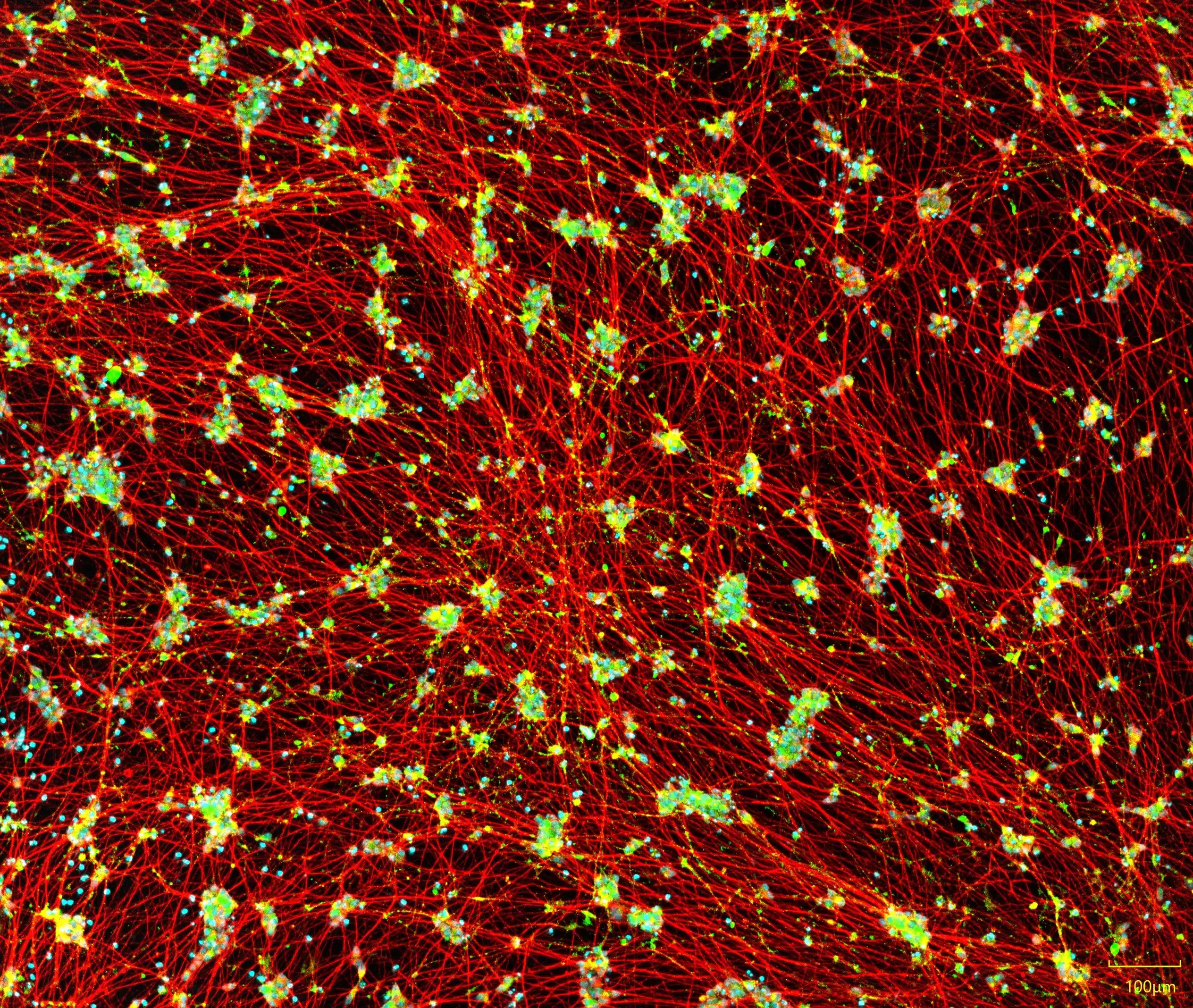
Excitatory Neuron Marker Expression
Expression of excitatory neuron–specific markers is evaluated post-differentiation to verify glutamatergic identity, purity, and maturation status. Using transcript and protein-level analyses, we confirm robust expression of established cortical and excitatory lineage markers to ensure batch-to-batch consistency and experimental reliability.
Merge

TUBB3

VGLUT1

Hoechst
iPSC-derived excitatory neurons express neuronal markers and display typical neurite growth. Immunofluorescent staining of Quick-Neuron™ Excitatory – Human iPSC-derived Neurons on Day 7 post-thaw that shows expression of the pan-neuronal marker TUBB3 and the glutamatergic neuron marker vGLUT1 (scale bar = 100 μm).
Functional Electrophysiology of Human iPSC-Derived Excitatory Neurons
Whole-Cell Patch Clamp

Electrophysiological properties of iPSC-derived excitatory neurons. Whole-cell patch clamp of Quick-Neuron™ Excitatory – Human iPSC-derived Neurons 5-6 weeks post-thaw. (A) A brightfield image of the neurons measured. (B) Spontaneous action potentials were recorded. (C) Spontaneous excitatory postsynaptic current of neurons was detected by voltage clamp measurement at -70mV, indicating the formation of mature synapses. Data courtesy of E-PHY SCIENCE SAS.
Multi-Electrode Array (MEA)
")
Excitatory Neurons seeded onto MEA plates remain functional after transport. An MEA plate seeded with Quick-Neuron™ Excitatory Neurons and primary human astrocytes was transported from Tokyo, Japan to Maryland, USA. The baseline network burst firing of one well was measured on day 49 post-thaw before transport (A) and on day 50 after 33-hr transport (B). (C) Network burst firing on day 51 exhibited responses to varying concentrations of 4-AP after transport. Data courtesy of Ricoh.
Transcriptomic Validation of Human iPSC-Derived Excitatory Neurons
RNA-Sequencing

Gene Expression of Quick-Neuron™ Excitatory Neurons. A heat map of selected gene expression data from RNA-seq performed on undifferentiated iPSCs, Quick-Neuron™ Excitatory Neurons (EX only) cultured for 10 days and 38 days, Quick-Neuron™ Excitatory Neurons cocultured with primary astrocytes (EX+AST) for 18 and 52 days, and primary fetal and adult brain samples is shown. Values represent log10(TPM + 1). Data courtesy of Ricoh.
Principal Component Analysis (PCA)
")
Quick-Neuron™ Excitatory Neurons display gene expression profiles similar to those of human brain: RNA-seq was performed on undifferentiated iPSCs, Quick-Neuron™Excitatory Neurons (EX only) cultured for 10 days and 38 days, Excitatory neurons cocultured with primary astrocytes (EX+AST) for 18 and 52 days, and primary fetal and adult brain samples. Principal component analysis indicates that Quick-Neuron™ Excitatory Neurons display gene expression similar to that of the human brain, particularly when grown with astrocytes. As cells remain in culture over time they more closely resemble adult human brain cells. Data courtesy of Ricoh.
Product Specifications
| Parameters | Specifications |
|---|---|
| Product Name | Quick-Neuron™ Excitatory Neurons - Human iPSC-Derived Excitatory Neurons |
| Catalog No. | EX-SeV-HC-CW50065 |
| Product Components | Cryopreserved cells, Component N, Component G2, and Component P |
| Starting Material | iPSCs derived from peripheral blood mononuclear cells (CIRM line CW50065) |
| Storage Conditions | Frozen cells should be stored in liquid nitrogen (vapor phase). The rest of the components should be stored at -20°C. |
| Cell Type | Excitatory Neurons (Glutamatergic) |
| Culture Type | Feeder Cell-Free |
| Disease | Healthy Donor |
| Donor Sex | Female |
| Donor Age at Sampling | 74 |
| Donor Race Ethnicity | Caucasian, not Latino |
| Patient History | See Resources for more information. |
| Reprogramming Method | Episomal vector |
| Induction Method | Transcription factors delivered by Sendai virus |
| Growth Properties | Adherent |
| Number of viable cells | > 1.0 million viable cells per vial upon thawing |
| Cell viability and remaining live cells |
>50% at day 1, >211 live cells per mm2 >50% at day 7, >211 live cells per mm2 |
| Differentiation |
At day 7 post-differentiation (CW50065) >80% TUBB3 positive cells >50% VGLUT1 positive cells among TUBB3 positive cells |
| Sterility | No growth observed |
| Mycoplasma | No mycoplasmal enzymes detected |
| Morphological Observation | Cells are adherent and neurites exhibit substantial outgrowth, elongation and branching, indicative of a differentiated phenotype. |
| Restricted Use | For research use only. Not for use in diagnostic or therapeutic procedures. |
Excitatory Neuron Resources
Episcopic Brightfield Imaging of Neuronal Cells on High-Density Microelectrode Arrays Enables Prediction of Cell Region Through AI Learning
Quick-Neuron™ Excitatory – Maintenance Medium
Quick-Neuron™ Excitatory – Human iPSC-Derived Neurons
Genetic and Functional Profiling of hiPSC-derived Excitatory Neurons Differentiated by Quick-Neuron™ Technology
Visualizing Neurite Outgrowth by Lentiviral Transduction of Fluorescent Proteins into Human iPSC-Derived Excitatory Neurons
No results found.
Related Products


FAQs
Do I need a license agreement for any of Ricoh Biosciences’ products?
No. You don’t need any licence or material transfer agreement (MTA) to use our differentiation kits or iPSC-derived cells. However, please be advised that these products are for research use only.
What kind of transcription factors are used for differentiation induction?
It is a proprietary formulated RNA and cannot be disclosed.
Does Quick-Tissue™ technology leave a genetic footprint?
Sendai virus (SeV) is an RNA virus, so it does not integrate into the genomic DNA. In principle, a foreign gene introduced intracellularly in the form of RNA is quickly translated and expressed because, unlike DNA, RNA does not need to enter the nucleus for forced expression, thereby providing no chance of mutagenesis. This is discussed in the following review paper: Yamamoto, et al., (2009) “Current prospects for mRNA gene delivery.” Eur. J. Pharm Biopharm 71, 484-489.
Will SeV remain active after differentiation?
No. The SeV used in our kits is a temperature-sensitive mutant that is active at 33℃ but becomes inactive at 37℃, which is the temperature instructed in the user guides post-differentiation.
No results found.
Contact Us
Have a question about our products, services, or custom projects? Our team is here to help—reach out and we’ll get back to you as soon as possible.
Subscribe
Sign up to our eNewsletter to stay up to date with the latest product launches, promotions, and receive expert tips.
By signing up you are agreeing to our Privacy Policy