

Amyotrophic Lateral Sclerosis (ALS)

Human iPSC-derived motor neuron models that recapitulate TDP-43 pathology and disease-relevant phenotypes for ALS drug discovery.
Human iPSC Models Built for ALS Research
Amyotrophic lateral sclerosis is a progressive neurodegenerative disease marked by motor neuron loss and hallmark pathologies like TDP-43 mislocalization, but traditional models often fail to capture this biology in a human-relevant way.
Ricoh Biosciences’ iPSC-derived motor neurons recapitulate key disease phenotypes across sporadic and familial ALS backgrounds, giving researchers a physiologically accurate, reproducible platform to study disease mechanisms and evaluate therapeutic candidates. From target validation to custom phenotypic assays, our team partners with you at every stage to generate translational data that moves ALS programs forward.
Why Choose iPSC-Derived Motor Neurons for ALS Models
Physiologically Relevant
ALS models recapitulate disease hallmarks including TDP-43 mislocalization and motor neuron degeneration.
Consistent & Reproducible
Produced under ISO-9001-certified processes to ensure robust performance and batch-to-batch consistency.
Sporadic & Familial Models
Patient-derived lines from sporadic cases as well as C9orf72, SOD1, and TARDBP mutations for broad disease coverage.
Naturally Evolving Pathology
ALS models exhibit progressive, spontaneous pathology that mirrors in-vivo disease and supports long-term studies.
Modeling TDP-43 Pathology: A Hallmark of ALS
A defining hallmark of ALS pathology is the mislocalization of TDP-43 protein from the nucleus to the cytoplasm, where it can form aggregates that disrupt RNA processing and contribute to motor neuron degeneration. Ricoh Biosciences’ iPSC-derived ALS motor neurons reproduce TDP-43 mislocalization, providing a powerful platform for evaluating therapeutic strategies targeting TDP-43 mislocalization and cytoplasmic accumulation.
TDP-43 Mislocalization Assay Workflow
Ricoh Biosciences’ TDP-43 mislocalization assay detects nuclear-to-cytoplasmic redistribution of TDP-43 in iPSC-derived motor neurons from a sporadic ALS donor, compared with a healthy donor control, just 8 days after thaw. The assay can be extended to additional ALS and control lines to capture donor-to-donor variability.
TDP-43 Mislocalization Assay Workflow. Schematic timeline of the TDP-43 mislocalization assay using Quick Neuron™ Motor – Human iPSC-Derived Neurons. Neurons are thawed and plated on Day 0, treated with vehicle or test compound on Day 7, and fixed, immunostained for TDP-43, and counterstained with Hoechst on Day 8.
Visualizing TDP-43 in ALS Motor Neurons
Immunofluorescence imaging of iPSC-derived motor neurons reveals distinct TDP-43 localization patterns in ALS versus healthy donor cultures. In healthy donor neurons, TDP-43 (green) is predominantly nuclear (cyan). In sporadic ALS neurons, TDP-43 is also detected in the cytoplasm, a defining pathological feature of ALS.
Edaravone restores nuclear TDP-43 localization in ALS motor neurons. Immunofluorescence of iPSC-derived motor neurons (Hoechst nuclear stain, cyan; TDP-43, green). Sporadic ALS neurons treated with vehicle (DMSO) show increased cytoplasmic TDP-43, a hallmark of ALS pathology. Edaravone treatment reduces cytoplasmic TDP-43, restoring a localization pattern similar to that of healthy donor neurons.


Edaravone Reduces Cytoplasmic TDP-43 Mislocalization
Edaravone treatment restores nuclear TDP-43 localization in ALS-derived motor neurons, significantly reducing pathological cytoplasmic mislocalization compared to DMSO control. Compound effects are assessed by comparing the ratio of cytoplasmic to total TDP-43 in treated cells with that of vehicle-treated cells. By combining accelerated differentiation with standardized imaging and analysis, this assay is designed to support fast, scalable studies, from pilot experiments to screening applications.
Edaravone corrects TDP-43 mislocalization in sporadic ALS motor neurons. Ratio of cytoplasmic to total TDP-43 in Quick Neuron™ iPSC-derived motor neurons. Healthy control neurons were treated with DMSO; sporadic ALS neurons were treated with DMSO or edaravone (10 µM or 100 µM) for 24 hours. Significance was quantified by pooled one-sided t-test: * = p < 0.05, ** = p < 0.01, *** = p < 0.001.

Visualizing ALS Model Development: From iPSCs to Functional Motor Neurons
Our ALS cell models begin with patient-derived iPSCs and mature into functional motor neurons with defined electrophysiological and morphological signatures. This standardized workflow ensures reproducible generation of iPSC motor neurons for ALS across studies and compound libraries.
Motor Neuron Differentiation Workflow
Our Quick Neuron™ workflow reliably generates high-quality motor neuron cultures within days, enabling scalable ALS phenotypic screening and biomarker discovery while maintaining consistency across lots.
Quick Neuron™ Motor – Human iPSC-Derived Neuron workflow. Schematic timeline of the Quick Neuron™ motor neuron differentiation and cryopreservation workflow.
Morphological Maturation and ALS Disease Phenotypes
During culture, ALS motor neuron models exhibit reduced survival and neurite complexity relative to healthy controls—phenotypes that emerge progressively and reflect the natural trajectory of ALS-associated neurodegeneration.


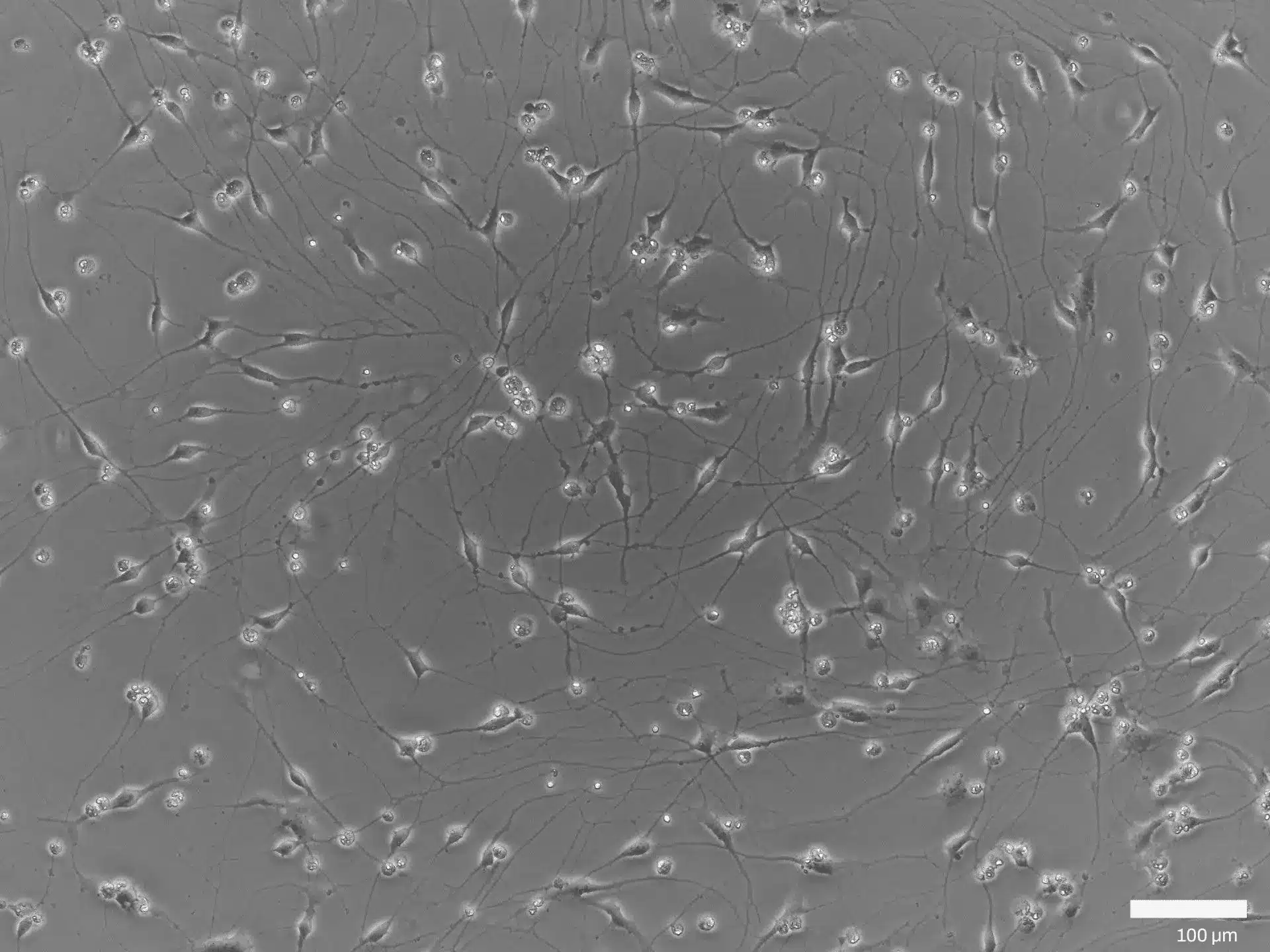

Morphology of Healthy vs. Sporadic ALS Motor Neurons. Representative phase contrast images of Quick-Neuron™ motor neurons derived from a sporadic ALS patient and a healthy control on days 1–7 post-thaw. (Scale bars, 100 µm).
Validation Through Marker Expression
Both ALS and control motor neurons express the pan-neuronal marker TUBB3 and motor neuron-specific markers ChAT and HB9, confirming lineage fidelity and successful differentiation. Combined with disease-specific phenotypes, these data validate our iPSC-derived ALS models as biologically accurate and reproducible platforms for ALS research.


Sporadic ALS and Control Motor Neurons Express Key Neuronal and Motor Neuron Markers. Immunofluorescence staining of Quick-Neuron™ motor neurons from a sporadic ALS patient and a healthy control. Cultures were stained for the pan-neuronal marker TUBB3 (red), the motor neuron-specific markers HB9 (green), ChAT (green), and nuclei (blue). (Scale bars, 100 µm).
ALS Disease Modeling Services for Drug Discovery & Preclinical Research
Accelerate ALS therapeutic programs using human-relevant ALS disease models purpose-built for neurodegeneration research. Ricoh Biosciences supports programs from early target evaluation through decision-enabling preclinical studies.
ALS-Relevant Cell Models
Deeply characterized human ALS models, including sporadic and familial ALS patient-derived motor neurons optimized for translational research.
Fee-for-Service
Assays
Custom ALS phenotypic assays using your compounds to generate high-quality, interpretable data in human motor neurons.
ALS Screening Partnerships
Long-term collaborations to evaluate compounds across disease-relevant phenotypes in ALS drug discovery models.
Preclinical Data Partnerships
Integrated generation of decision-enabling preclinical ALS cell model data packages to support therapeutic advancement.
Request a Free Consultation
Work directly with our scientific team to evaluate feasibility, timelines, scale, and validation strategies for your program.
Online Marketplaces
Our products and services are available through Science Exchange and Scientist.com for efficient, compliant procurement with existing vendor frameworks.
"*" indicates required fields