

Quick Neuron™ Sensory – mRNA Kit
Quick-Neuron™ Sensory – mRNA Kits enable the differentiation of human pluripotent stem cells into sensory neurons using mRNA-based technology through an optimized and reproducible workflow.
Note: This kit is no longer available for individual sale. We now offer bulk orders and custom differentiation services to support your project needs.
Advantages of iPSC-Derived Sensory Neurons
Sensory Neuron Differentiation Kit Protocol
Explore our detailed differentiation protocols, a step-by-step guide designed to simplify and optimize your laboratory procedures using our iPSC-derived cells and differentiation kits. These protocols leverage the latest advancements in iPSC technology to ensure efficient and reproducible results.


Sensory Neuron Characterization
Characterization of iPSC-derived sensory neurons is crucial to ensure their utility in research. Employing sensory neuron markers, we can confirm the identity and purity of these neurons.
Sensory Neuron Marker Expression
Understanding the role of sensory neuron markers is crucial in neuroscience research. Our comprehensive guide delves into the identification and significance of these markers in iPSC-derived neurons, providing essential information for researchers.

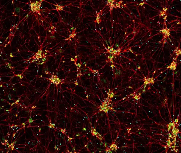
iPSC-derived sensory neurons express neuronal markers and display typical neurite growth. Immunofluorescent staining of Quick-Neuron™ Sensory – mRNA Kit cell cultures shows typical neurite growth and expression of PRPH and BRN3A on day 10 post-differentiation (scale bar = 100 μm). Staining conditions: Peripherin Rabbit polyclonal antibody (VI3079111) and BRN3A(14A6):sc-8429 mouse (B0521), 1:100, 1:2000. Nuclei were counterstained with Hoechst 33342.
Real-time Quantitative PCR Analysis
Real-time quantitative PCR analysis of expression levels of sensory neuron-associated genes SCN9A, PRPH, ISL1, and BRN3A were examined. The graph shows comparison of gene expression in Quick-Neuron™ – Sensory mRNA Culture on day 10 with gene expression in Human Dorsal Root Ganglion total RNA (TaKaRa, Catalog Number: 636150). The relative gene expression is normalized to phosphoglycerate kinase 1 (PGK1), and then calculated as a fold induction relative to hiPSCs as a control. Error bars show standard deviation.

Product Specifications
| Parameters | Specifications |
|---|---|
| Product Name | Quick-Neuron™ Sensory – mRNA Kit |
| Catalog No. | SS-mRNA |
| Product Components | QNS-mRNA-P, Component N, Component S1, and Component P |
| Storage Conditions | mRNA should be stored at -80°C. All other components can be stored at -20°C or -80°C. |
| Cell Type | Sensory Neurons |
| Induction Method | Transcription factors delivered by synthetic mRNA |
| Differentiation |
At Day 7 Post-Differentiation: >80% TUBB3+, >50% PRPH+, >50% SCN9A+/TUBB3+ (Stain for ISL1, no criteria) |
| Sterility | No growth observed |
| Mycoplasma | No DNA detected |
| Restricted Use | For research use only. Not for use in diagnostic or therapeutic procedures. |