
Quick Glia™ Astrocyte – SeV Kit
Quick-Glia™ Astrocyte – SeV Kits enable the differentiation of human pluripotent stem cells into astrocytes using Sendai virus technology through a streamlined and reproducible workflow.
Note: This kit is no longer available for individual sale. We now offer bulk orders and custom differentiation services to support your research and development needs.
Advantages of iPSC-Derived Astrocytes
Astrocyte Differentiation Kit Protocol
Our optimized astrocyte differentiation protocol supports reproducible generation of GFAP+, ALDH1L1+ astrocytes from human induced pluripotent stem cells (iPSCs) over a ~4-week timeline. This step-by-step workflow outlines the key culture stages and media changes required to generate functionally mature, highly pure astrocyte cultures for neuroinflammation research, co-culture studies, and disease modeling applications.
 Representative workflow for Quick-Glia™ Astrocyte – SeV Kit

Astrocyte Characterization
Astrocytes are the most abundant glial cell type in the CNS, supporting neuronal metabolism, synapse formation, and blood-brain barrier integrity, and playing a central role in neuroinflammation and neurodegenerative disease. Confirming a clean astrocytic phenotype at every lot is essential before these cells can be trusted in co-culture, disease modeling, or drug screening applications.
Astrocyte Marker Expression
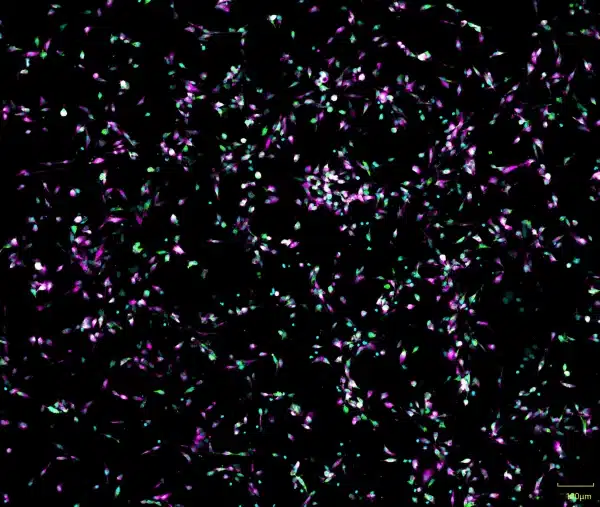
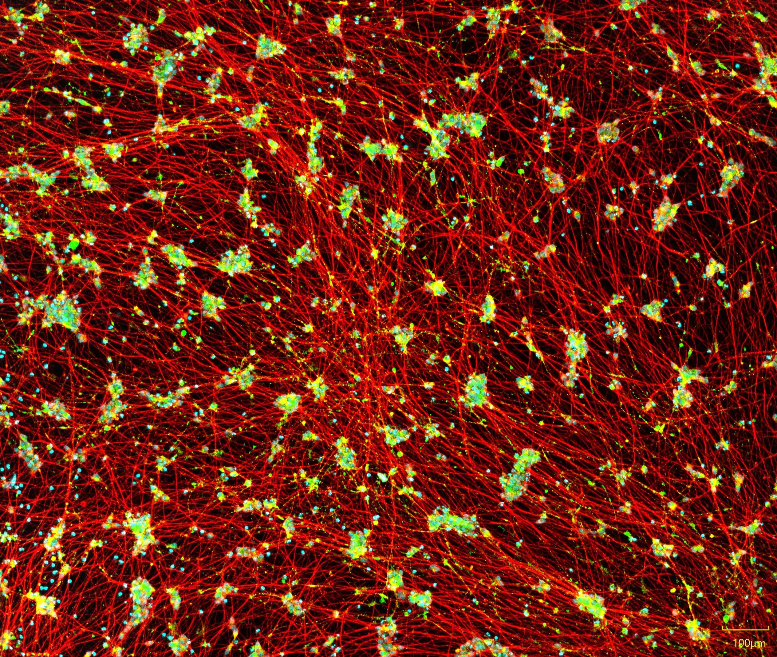
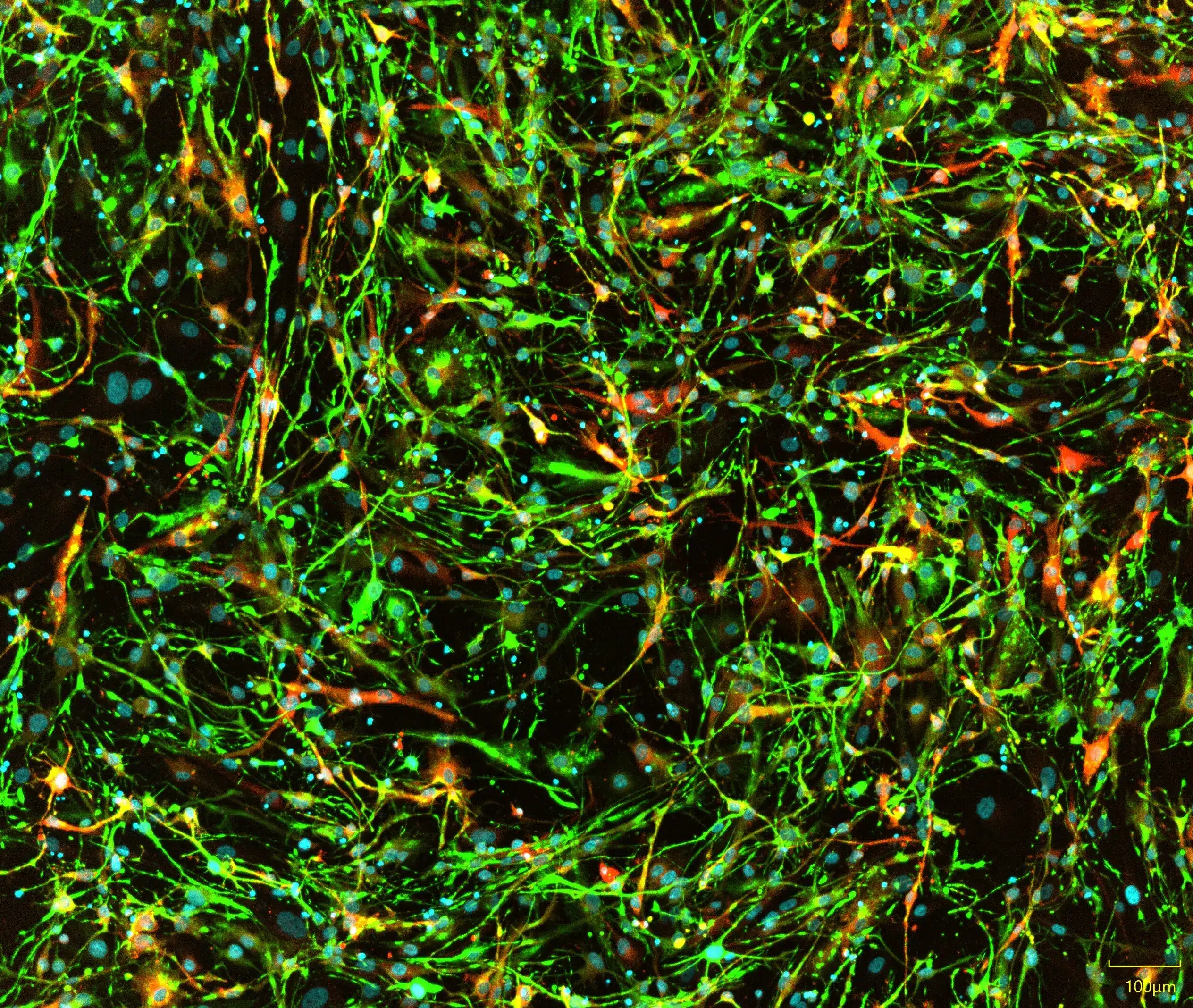
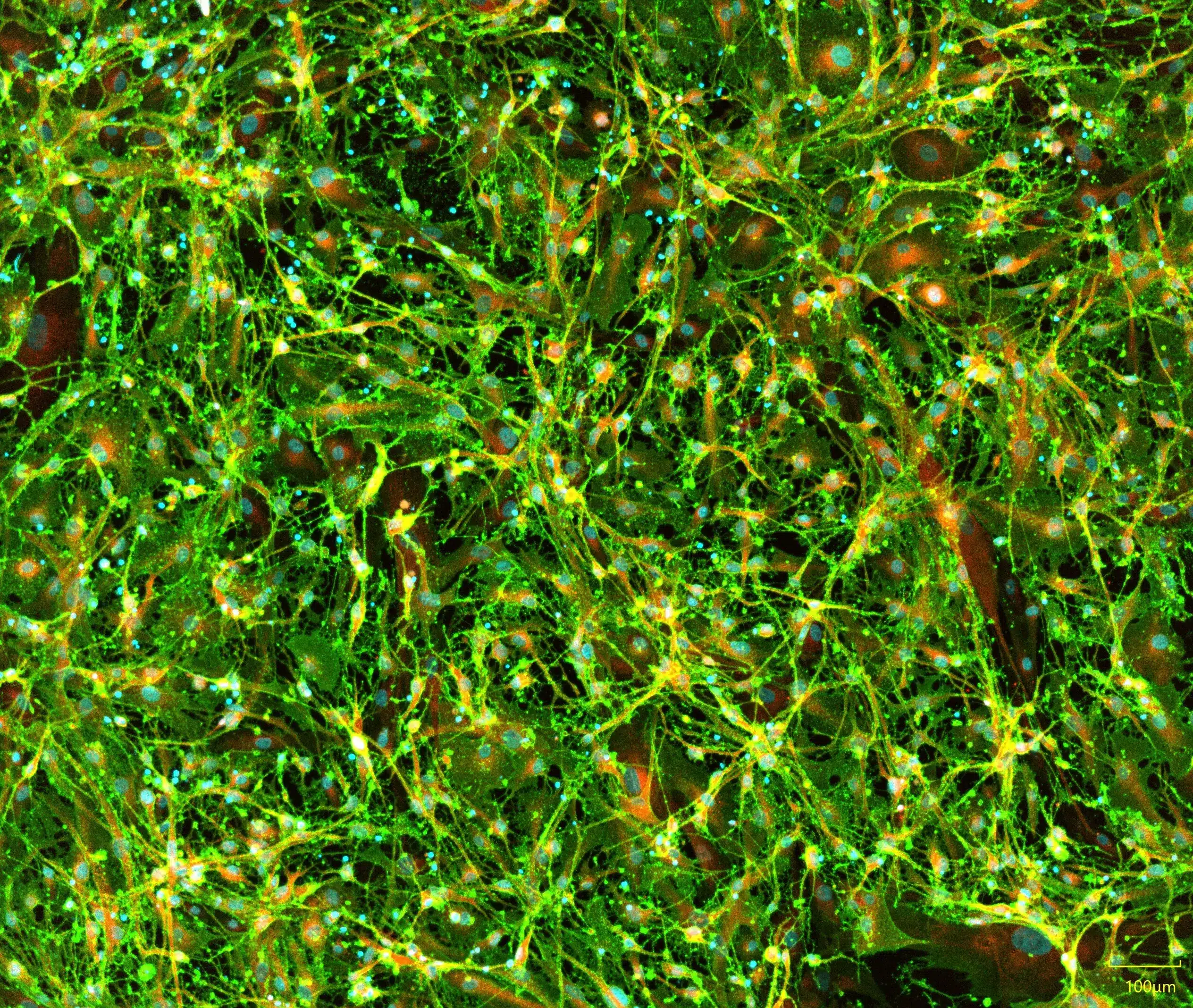
Quick-Glia™ Astrocytes are characterized by immunocytochemistry for a panel of canonical astrocyte markers. GFAP, an intermediate filament protein, confirms astrocytic cytoskeletal identity, while ALDH1L1 and CD44 mark mature, pan-reactive astrocyte populations, together supporting confidence in cell identity and purity from the moment of thaw.
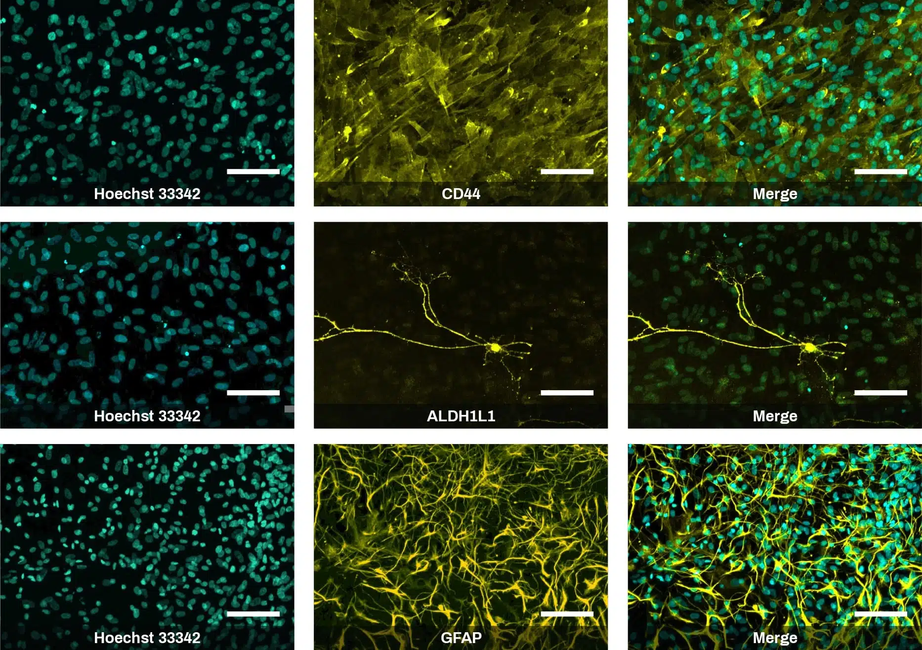
iPSC-derived astrocytes express glial markers and display typical astrocytic morphology. Immunofluorescent staining of Quick-Glia™ Astrocyte shows expression of astrocytic markers CD44, ALDH1L1, and GFAP. Nuclei are counterstained with Hoechst 33342 (cyan) (scale bars = 100 μm).
Transcriptomic Similarity to Primary Astrocytes

Quick-Glia™ Astrocytes closely resemble the transcriptomic profile of primary human astrocytes. (A) Gene expression profiles of iPSCs and Quick-Glia™ – Astrocyte SeV Culture on day 28 were compared with the profile of human primary astrocytes and the results are shown as scatter plots. The horizontal axis indicates the expression levels of genes in human primary astrocytes purchased from ScienCell (Catalog Number: 1800-5), whereas the vertical axis indicates the expression levels of genes in iPSCs (left) and in Quick-Glia™ – Astrocyte SeV Culture on day 28 (right). The levels of gene expression are shown based on transcripts per million (TPM) in the log10 scale. Blue and green dots represent upregulated and downregulated genes (FDR<0.05), respectively, relative to their levels in human primary astrocytes. (B) Similarities of gene expression profiles of human iPSCs and Quick-Glia™ – Astrocyte SeV Culture on days 14, 28 and 42 to the profile of human primary astrocytes are shown as a bar chart. The vertical axis indicates Pearson correlation (r) based on median-subtracted logTPM.
Quantitative PCR Analysis

Real-time quantitative PCR analysis of expression levels of astrocyte-associated genes CD44, GFAP, S100β and ALDH1L1. Graphs show comparison of Quick-Glia™ – Astrocyte SeV Culture on day 28 (A) and day 42 (B) with human brain total RNA (TaKaRa, Catalog Number: 636530). The relative gene expression is normalized to phosphoglycerate kinase 1 (PGK1), and then calculated as a fold induction relative to undifferentiated hPSCs as a control. Error bars show standard deviation.
Functional Validation of iPSC-Derived Astrocytes
Glutamate Uptake Assay
Glutamate clearance from the extracellular space is a core astrocytic function, protecting neurons from excitotoxicity and supporting healthy synaptic signaling. Quick-Glia™ Astrocytes are functionally validated for glutamate uptake alongside human primary astrocytes, confirming that these cells perform this essential physiological role and are suitable for use in excitotoxicity, synaptic function, and neuroinflammation research.

Glutamate uptake assay. Glutamate clearance by Quick-Glia – Astrocyte SeV kit cultures harvested at 28 or 42 days post-infection, compared with human primary astrocytes and human iPSCs (negative control). Cells were incubated with 100 µM L-glutamate for 60 minutes, and the remaining glutamate was measured using the Glutamate-Glo assay (Promega) and normalized to live cell count.
Product Specifications
| Parameters | Specifications |
|---|---|
| Product Name | Quick-Glia™ Astrocyte - SeV Kit |
| Catalog No. | AS-SeV |
| Product Components | QGA-SeV, Component N1, Component GA1, Component GA2, and Coating Material A |
| Storage Conditions | SeV should be stored at -80°C. All other components can be stored at -20°C or -80°C. |
| Cell Type | Astrocytes |
| Induction Method | Transcription factors delivered by Sendai virus |
| Differentiation | Log10 RQ Value: ALDH1L1 (>1), GFAP (>4), CD44 (>1.5), SLC1A3 (>1.5) |
| Sterility | No growth observed |
| Mycoplasma | No DNA detected |
| Restricted Use | For research use only. Not for use in diagnostic or therapeutic procedures. |