
Download Your Application Note
"*" indicates required fields
We respect your inbox. You can unsubscribe at any time. View our privacy policy.
Inside this application note
Review lentiviral transduction conditions, optimal MOI data, and live-cell imaging results for neurite outgrowth visualization, glutamate excitotoxicity, and GCaMP6f-based calcium oscillation monitoring in hiPSC-derived excitatory neurons.
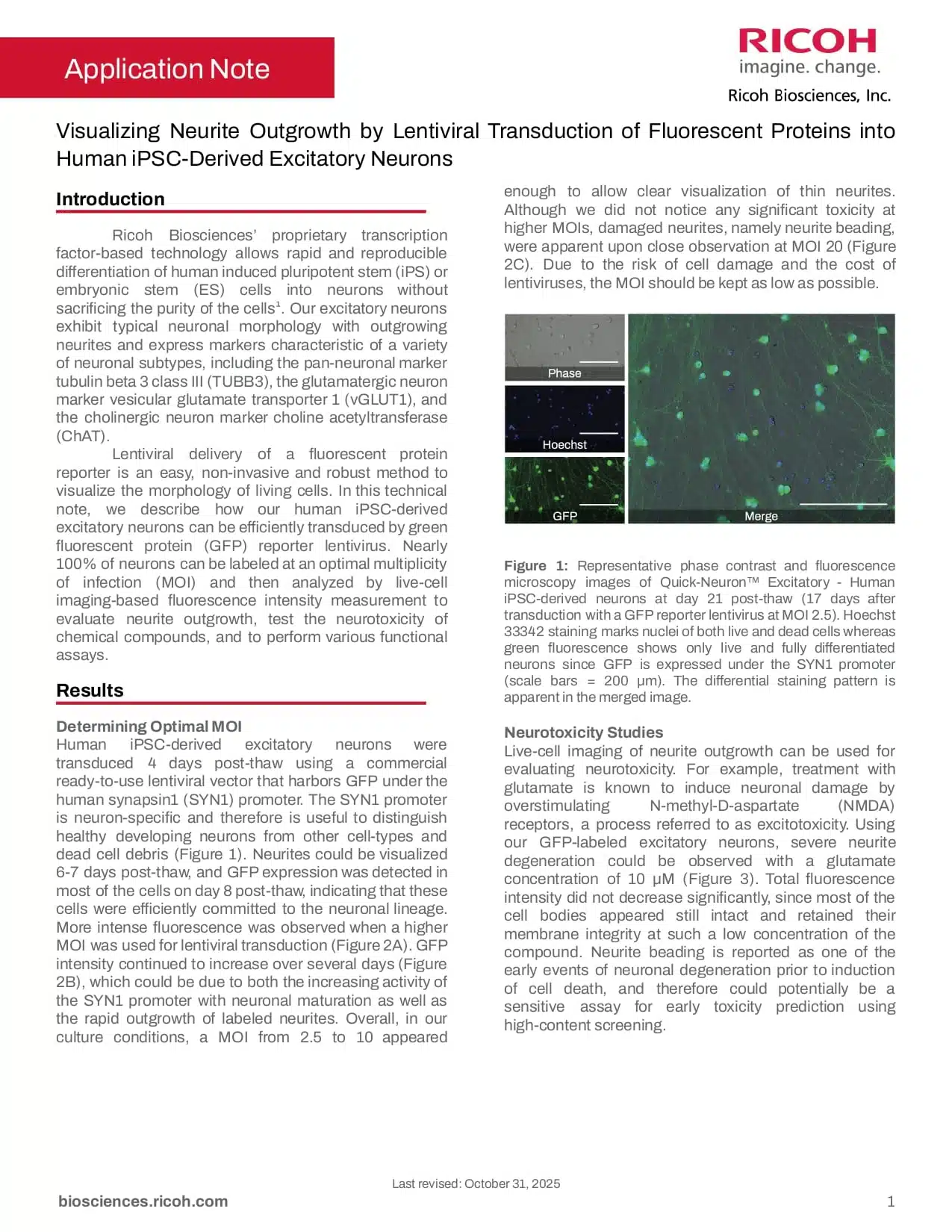
- GFP expression detected in the majority of cells by day 8 post-thaw using a SYN1 promoter-driven lentiviral vector, with MOI 2.5–10 sufficient for clear visualization of thin neurites without significant toxicity
- Severe neurite degeneration observed at 10 μM glutamate by day 21 post-thaw, with neurite beading identified as a potential sensitive early endpoint for neurotoxicity prediction prior to cell death
- Spontaneous calcium oscillations detected from day 21 post-thaw using GCaMP6f, with the most stable signals at approximately 1 peak per minute obtained at day 29 post-thaw at MOI 30
Related Resources
Explore related cell models, services, and protocols connected to this application.