Quick-Neuron™ Motor – Human iPSC-Derived Neurons (Healthy Donor)

Quick-Neuron™ Motor – Human iPSC-Derived Neurons (Healthy Donor)
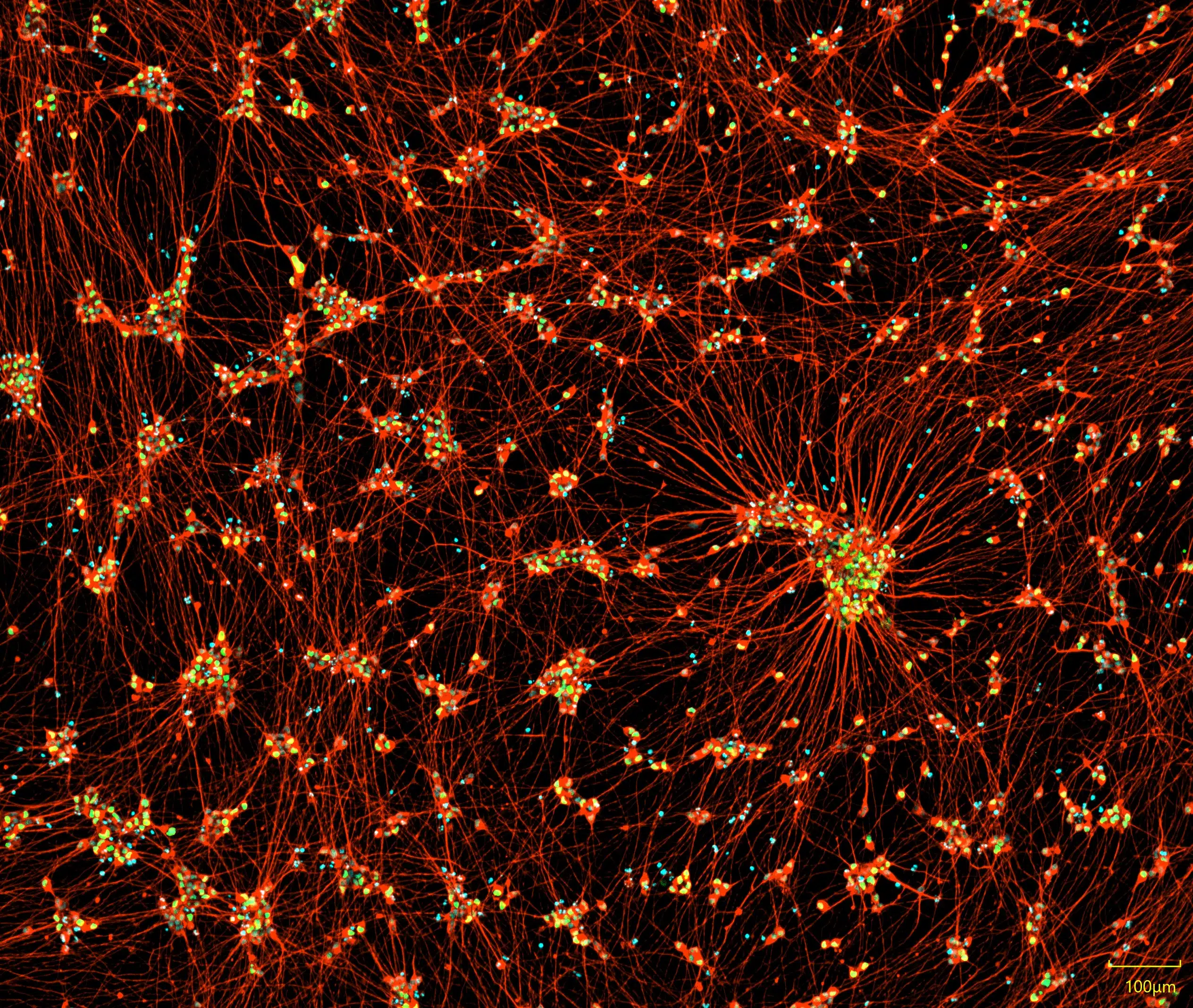
Our proprietary transcription factor-based stem cell differentiation method produces neurons without a genetic footprint. Quick-Neuron™ Motor – Human iPSC-derived Neurons display typical neurite outgrowth and express a variety of neuronal markers, such as the pan-neuronal marker TUBB3 (red) and the motor neuron marker HB9 (green). When thawed and maintained according to the instructions in the user guide, the iPSC-derived neurons are viable long-term and are suitable for a variety of characterization and neurotoxicity assays.
$850.00
Advantages of iPSC-Derived Motor Neurons
~ 1 Week Differentiation
Functionally Validated
Highly Pure Population
No Genetic Footprint
Motor Neuron Protocol
Explore our detailed differentiation protocols, a step-by-step guide designed to simplify and optimize your laboratory procedures using our iPSC-derived cells and differentiation kits. These protocols leverage the latest advancements in iPSC technology to ensure efficient and reproducible results.


Motor neuron morphology is confirmed via phase contrast imagery: Representative images of Quick-Neuron™ Motor – Human iPSC-derived Neurons on days 1-7 post-thaw (scale bar = 100 μm).
Motor Neuron Characterization
Characterization of iPSC-derived motor neurons is crucial to ensure their utility in research. Employing motor neuron markers, we can confirm the identity and purity of these neurons.
Motor Neuron Marker Expression
Understanding the role of motor neuron markers is crucial in neuroscience research. Our comprehensive guide delves into the identification and significance of these markers in iPSC-derived neurons, providing essential information for researchers.
Merge

TUBB3

HB9

Nuclei
iPSC-derived motor neurons express neuronal markers and display typical neurite outgrowth. Immunofluorescent staining of iPSC-derived motor neurons at day 10 post-differentiation. Cells exhibit extensive neurite outgrowth and co-expression of the pan-neuronal marker TUBB3 and the motor neuron-specific marker HB9.
Real-time Quantitative PCR Analysis

Real-time quantitative PCR analysis of expression levels of genes CHAT, HB9, and ISL1 were examined. The graph shows gene expression in Quick-Neuron™ Motor – SeV culture on day 10 post-differentiation. The relative gene expression is normalized to phosphoglycerate kinase 1 (PGK1), and then calculated as a fold induction relative to undifferentiated hPSCs as a control. Error bars show standard deviation.
TDP-43 Mislocalization in ALS Motor Neuron Models
TDP-43 mislocalization is a defining molecular feature of amyotrophic lateral sclerosis (ALS). Our ALS motor neuron models enable detailed investigation of TDP-43 biology and support a range of applications in disease mechanism studies and therapeutic discovery.
Quantification of TDP-43 localization in Quick-Neuron™ motor neurons. Violin plots show the ratio of cytoplasmic to total TDP-43 puncta in cultures derived from a sporadic ALS patient and a healthy control. Significance was quantified by T-test. Stars denote statistical significance: **** = p <0.0001.

Product Specifications
| Parameters | Specifications |
|---|---|
| Product Name | Quick-Neuron™ Motor - Human iPSC-Derived Neurons |
| Catalog No. | MT-SeV-HC-CW50065 |
| Product Components | Cryopreserved cells, Component N1, Component A, Component P, and Component K |
| Starting Material | iPSCs derived from peripheral blood mononuclear cells (CIRM line CW50065) |
| Storage Conditions | Frozen cells should be stored in liquid nitrogen (vapor phase). The rest of the components should be stored at -20°C. |
| Cell Type | Motor Neurons |
| Culture Type | Feeder Cell-Free |
| Disease | Healthy Control |
| Donor Sex | Female |
| Donor Age At Sampling | 74 |
| Donor Race Ethnicity | Caucasian, not Latino |
| Patient History | See Resources section for more information. |
| Reprogramming Method | Episomal vector |
| Induction Method | Transcription factors delivered by Sendai virus |
| Growth Properties | Adherent |
| Number of viable cells | > 1.0 million viable cells per vial upon thawing |
| Cell viability and remaining live cells | >50% at day 1, >211 live cells per mm2 >50% at day 7, >211 live cells per mm2 |
| Differentiation | >80% TUBB3 positive cells >50% ChAT positive cells among TUBB3 positive cells >40% HB9 positive cells among TUBB3 positive cells |
| Sterility | No growth observed for Bacteria and Fungus |
| Mycoplasma | No contamination detected |
| Morphological Observation | Cells are adherent and neurites exhibit substantial outgrowth, elongation and branching, indicative of a differentiated phenotype. |
| Restricted Use | For Research Use Only. Not for use in diagnostic or therapeutic procedures. |
Resources
Quick-Neuron™ Motor – Human iPSC-Derived Neurons
Quick-Neuron™ Motor – SeV Kit
Human Motor Neurons With SOD1-G93A Mutation Generated From CRISPR/Cas9 Gene-Edited iPSCs Develop Pathological Features of Amyotrophic Lateral Sclerosis.
Build Disease Models That Matter: The Power of Custom iPSC Differentiation
Modeling ALS Using Patient-Derived iPSCs: A Human-Relevant Platform for Disease Research and Therapeutic Discovery (SfN 2025)
No results found.
Related Products
Related products

FAQs
Does Quick-Tissue™ technology leave a genetic footprint?
Sendai virus (SeV) is an RNA virus, so it does not integrate into the genomic DNA. In principle, a foreign gene introduced intracellularly in the form of RNA is quickly translated and expressed because, unlike DNA, RNA does not need to enter the nucleus for forced expression, thereby providing no chance of mutagenesis. This is discussed in the following review paper: Yamamoto, et al., (2009) “Current prospects for mRNA gene delivery.” Eur. J. Pharm Biopharm 71, 484-489.
Will SeV remain active after differentiation?
No. The SeV used in our kits is a temperature-sensitive mutant that is active at 33℃ but becomes inactive at 37℃, which is the temperature instructed in the user guides post-differentiation.
Is Sendai virus (SeV) dangerous?
SeV is not pathogenic to humans (i.e., humans are not the natural host of the virus) and the infection does not persist in immunocompetent animals. Furthermore, SeV used in our kits does not produce infectious viral particles upon transduction to host hPSCs and is a temperature-sensitive mutant, such that it is active at 33℃ but becomes inactive at 37℃. However, because SeV can be transmitted by aerosol and contact with respiratory secretions and is highly contagious, appropriate care must be taken to prevent potential mucosal exposure to the virus. Our SeV-based kits must be used under Biosafety Level 2 (BL-2) containment with a biological safety cabinet or a laminar flow hood and with appropriate personal protective equipment. In the event that the virus comes into contact with skin or eyes, decontaminate the affected area by flushing with plenty of water and follow the safety manual prepared by your laboratory and approved by your Institutional Biosafety Committee.
Do I need a license agreement for any of Ricoh Biosciences’ products?
No. You don’t need any licence or material transfer agreement (MTA) to use our differentiation kits or iPSC-derived cells. However, please be advised that these products are for research use only.
What kind of transcription factors are used for differentiation induction?
It is a proprietary formulated RNA and cannot be disclosed.
No results found.
Contact Us
Have a question about our products, services, or custom projects? Our team is here to help—reach out and we’ll get back to you as soon as possible.
Subscribe
Sign up to our eNewsletter to stay up to date with the latest product launches, promotions, and receive expert tips.
By signing up you are agreeing to our Privacy Policy