

Quick-Neuron™ Cholinergic – SeV Kit
This kit differentiates human pluripotent stem cells into cholinergic neurons in 6 days using Sendai virus.
Note: This kit is no longer available for individual sale. We now offer bulk orders and custom differentiation services.
Advantages of iPSC-Derived Cholinergic Neurons
~ 1 Week Differentiation
Optimized & Reproducible
Highly Pure Population
No Genetic Footprint
Patient-Derived Cholinergic Neurons
We offer cholinergic neurons derived from human iPSCs licensed through The California Institute for Regenerative Medicine (CIRM). These neurons have a diverse genetic background, as hiPSC lines from over 1,500 donors are available in the CIRM repository. If you require neurons from a specific donor, contact us to discuss your needs.
Cholinergic Neuron Differentiation Kit Protocol
Explore our detailed differentiation protocols, a step-by-step guide designed to simplify and optimize your laboratory procedures using our iPSC-derived cells and differentiation kits. These protocols leverage the latest advancements in iPSC technology to ensure efficient and reproducible results.


Cholinergic neuron morphology is confirmed via phase contrast imagery: Representative images of Quick-Neuron™ Cholinergic – SeV Kit cell cultures on days 1-6 post-differentiation (scale bar = 100 μm).
Cholinergic Neuron Characterization
Characterization of iPSC-derived cholinergic neurons is crucial to ensure their utility in research. Employing cholinergic neuron markers, we can confirm the identity and purity of these neurons.
Cholinergic Neuron Marker Expression
Understanding the role of cholinergic neuron markers is crucial in neuroscience research. Our comprehensive guide delves into the identification and significance of these markers in iPSC-derived neurons, providing essential information for researchers.

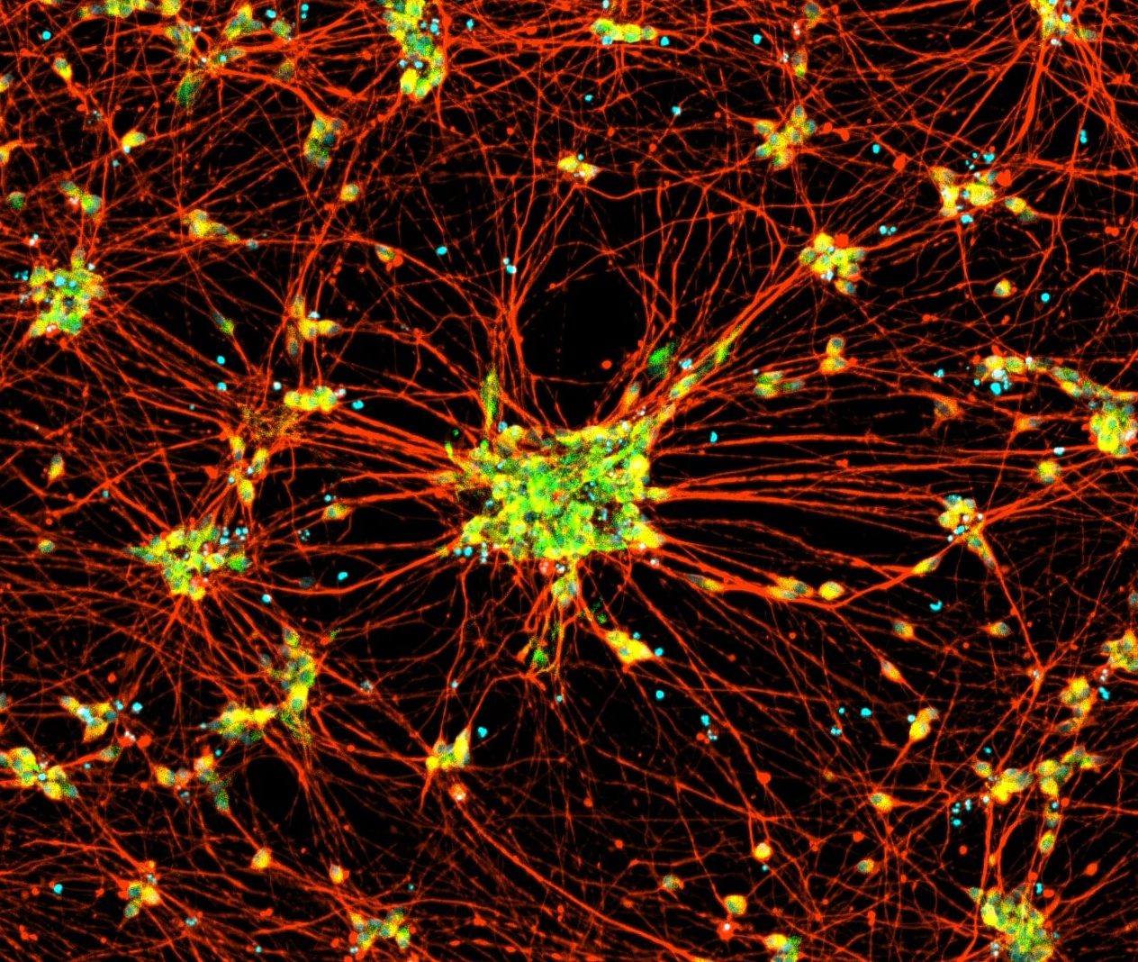
iPSC-derived cholinergic neurons express neuronal markers and display typical neurite growth. Immunofluorescent staining of Quick-Neuron™ Cholinergic – Human iPSC-derived Neurons on Day 3 post-thaw that shows expression of the pan-neuronal marker TUBB3 and the cholinergic neuron marker ChAT (scale bar = 100 μm).
Additional Cholinergic Neuron Data
ALS Disease Study

ALS Disease Study: Healthy control and ALS patient-derived iPSCs were differentiated into cholinergic neurons. The cells were fixed at day 10 from iPSCs and stained by TUBB3 (Green) and TDP-43 (Red).
Neurite Length Analysis

Neurite Length Analysis: Our fast differentiation enables quicker and quantitative comparison between healthy and patient or treated and non-treated cultures..
Product Specifications
| Parameters | Specifications |
|---|---|
| Product Name | Quick-Neuron™ Cholinergic - SeV Kit |
| Catalog No. | CH-SeV |
| Product Components | QN-SeV-P, Component N1, Component A, Component P, and Coating Material A |
| Storage Conditions | SeV should be stored at -80°C. All other components can be stored at -20°C or -80°C. |
| Cell Type | Cholinergic Neurons |
| Induction Method | Transcription factors delivered by Sendai virus |
| Differentiation | At day 7 post-differentiation (CW50065): >80% TUBB3+, >50% ChAT+/TUBB3+ |
| Sterility | No growth observed |
| Mycoplasma | No DNA detected |
| Restricted Use | For Research Use Only. Not for use in diagnostic or therapeutic procedures. |
Resources
Quick-Neuron™ Cholinergic – Human iPSC-Derived Neurons
Quick-Neuron™ Cholinergic – SeV Kit
Induction of specific neuron types by overexpression of single transcription factors.
Protective effects of NAMPT or MAPK inhibitors and NaR on Wallerian degeneration of mammalian axons
Tools to Study Neurodegeneration: iPSC-derived Neurons, Nerve Organoids, and Synaptogenic Beads
No results found.
FAQs
What kind of transcription factors are used for differentiation induction?
It is a proprietary formulated RNA and cannot be disclosed.
Why do I see many floating cells and/or cellular debris on day 1?
hPSCs were likely damaged while they were harvested. hPSCs may have been mechanically harvested by scraping or were pipetted too much or too vigorously. Lift Solution D1-treated hPSCs only by pipetting them up and down up to 15 times. The mixing of hPSCs with SeV may have also been done too strongly. Gently rock the plate several times to mix hPSCs with SeV.
What should I do if I find on day 1 that cells were accumulated in the center of the well?
It is not recommended to start mRNA-based differentiation induction because the differentiation efficiency is expected to be quite low. For successful differentiation using our mRNA-based methods including Mesendoderm Booster, hPSCs should be evenly distributed in the well. It is likely that the plate was shaken too much, and cells were accumulated in the center of the well while they were floating. Next time, please handle the plate more gently or it can be left on a flat bench top without vibration for 15 min before it is put inside a CO₂ incubator.
What should I do if I find on day 1 that my culture was seeded only in the center but not in the periphery of the well?
Unless cells are accumulated in the center of the well, the instructions can be followed since this is an issue of coating. We have observed that 4-well plates available from Nunc sometimes do not allow cells to settle down in the periphery of the well when our coating conditions are applied. Next time, please use a 24-well plate and try the following: 1) incubate the plate with Coating Material A at 4°C overnight instead of 37°C for 2 hours, 2) do not aspirate the supernatant completely or leave the supernatant just enough to cover the well bottom surface after coating with Coating Material A is completed and 3) do not touch the bottom of wells with fingers when handling the plate.
I plated cells as instructed, so why does my culture look less confluent than the one shown in the user guide on day 1?
hPSCs were likely damaged while they were harvested. Ideally, hPSCs should be lifted only by pipetting several times using Solution D1 as instructed in the user guide. Even if pipetting 15 times does not lift all of the cells in the culture, do not attempt to do more pipetting but collect only cells that come off. Instead rinse the culture with PBS and start Solution D1 treatment again but with longer incubation (up to 20 minutes). Next time please reduce the concentration of the substrate used to coat the dish/well for the maintenance of the source hPSCs to 50-70%.
Why does my culture have colonies that appear balled up and round on day 1?
It could be that Coating Material A was diluted at room temperature or diluted using PBS warmed to room temperature. Keep both Coating Material A and PBS on ice before dilution.
Can I use an orbital shaker for SeV infection while incubating cell suspension for 10 minutes?
The purpose of flicking the tube containing cells and SeV every 2 minutes during the 10 minute incubation is to prevent cells from settling down and forming aggregates. If the orbital shaker can do so, then it is possible to use it.
Can I use Geltrex or Matrigel as a coating substrate for hPSC cultures?
Our kits are not optimized with these substrates. However, if your hPSC differentiation protocols are already optimized with such a substrate, you may try using one of these with our kit at your own risk.
I was able to harvest more hPSCs than I needed for the kit. Can I replate or freeze the leftover hPSCs to maintain their culture?
No. If you’re following the instructions given in the user guide, the hPSCs will be harvested in a differentiation medium so the leftover hPSCs cannot be maintained.
Why are there many aggregates after harvesting hPSCs?
After harvesting hPSCs, cells should not be left in the collection tube for more than 5 minutes. Leaving cells for a longer period may cause the formation of cellular clumps which interferes with infection. Single cells are best for infection.
Why doesn’t Solution D1 dissociate my hPSC culture without scraping?
The concentration of the substrate used to coat the dish/well was likely too high, so Solution D1 treatment requires longer incubation than 10 minutes (up to 20 minutes). Next time, please reduce the concentration of the substrate to 50-70%.
Why do differentiation kits require hPSCs to be maintained under StemFit or StemFlex conditions?
hPSCs should be adapted to passaging as single cells prior to differentiation. Our protocols have only been tested with StemFit/StemFlex, so we can’t guarantee results using other culture conditions.
How long do I need to maintain hPSCs prior to beginning to use your kit?
Before beginning differentiation, cultures should be healthy and displaying typical morphologies (i.e., round colonies with tightly packed cells exhibiting a small cytoplasmic area), few spontaneously differentiating cells, and normal growth rates (i.e., doubling once a day). We recommend growing the cells for at least 14 days post-thaw before differentiating and not allowing the cultures to reach more than 80% confluency.
Do I need a license agreement for any of Ricoh Biosciences’ products?
No. You don’t need any licence or material transfer agreement (MTA) to use our differentiation kits or iPSC-derived cells. However, please be advised that these products are for research use only.
Is Sendai virus (SeV) dangerous?
SeV is not pathogenic to humans (i.e., humans are not the natural host of the virus) and the infection does not persist in immunocompetent animals. Furthermore, SeV used in our kits does not produce infectious viral particles upon transduction to host hPSCs and is a temperature-sensitive mutant, such that it is active at 33℃ but becomes inactive at 37℃. However, because SeV can be transmitted by aerosol and contact with respiratory secretions and is highly contagious, appropriate care must be taken to prevent potential mucosal exposure to the virus. Our SeV-based kits must be used under Biosafety Level 2 (BL-2) containment with a biological safety cabinet or a laminar flow hood and with appropriate personal protective equipment. In the event that the virus comes into contact with skin or eyes, decontaminate the affected area by flushing with plenty of water and follow the safety manual prepared by your laboratory and approved by your Institutional Biosafety Committee.
Will SeV remain active after differentiation?
No. The SeV used in our kits is a temperature-sensitive mutant that is active at 33℃ but becomes inactive at 37℃, which is the temperature instructed in the user guides post-differentiation.
Does Quick-Tissue™ technology leave a genetic footprint?
Sendai virus (SeV) is an RNA virus, so it does not integrate into the genomic DNA. In principle, a foreign gene introduced intracellularly in the form of RNA is quickly translated and expressed because, unlike DNA, RNA does not need to enter the nucleus for forced expression, thereby providing no chance of mutagenesis. This is discussed in the following review paper: Yamamoto, et al., (2009) “Current prospects for mRNA gene delivery.” Eur. J. Pharm Biopharm 71, 484-489.
No results found.
Contact Us
Have a question about our products, services, or custom projects? Our team is here to help—reach out and we’ll get back to you as soon as possible.
Subscribe
Sign up to our eNewsletter to stay up to date with the latest product launches, promotions, and receive expert tips.
By signing up you are agreeing to our Privacy Policy