Quick-Neuron™ GABAergic – mRNA Kit
Quick-Neuron™ GABAergic – mRNA Kit
This kit differentiates human pluripotent stem cells into GABAergic neurons in 10 days using synthetic mRNA.
Advantages of iPSC-Derived GABAergic Neurons
~ 1 Week Differentiation
Optimized & Reproducible
Highly Pure Population
No Genetic Footprint
GABAergic Neuron Differentiation Kit Protocol
Explore our detailed differentiation protocols, a step-by-step guide designed to simplify and optimize your laboratory procedures using our iPSC-derived cells and differentiation kits. These protocols leverage the latest advancements in iPSC technology to ensure efficient and reproducible results.


GABAergic neuron morphology is confirmed via phase contrast imagery: Representative images of Quick-Neuron™ GABAergic – mRNA Kit cell cultures on days 1-10 post-differentiation (scale bar = 100 μm).
GABAergic Neuron Characterization
Characterization of iPSC-derived GABAergic neurons is crucial to ensure their utility in research. Employing GABAergic neuron markers, we can confirm the identity and purity of these neurons.
GABAergic Neuron Marker Expression
Understanding the role of GABAergic neuron markers is crucial in neuroscience research. Our comprehensive guide delves into the identification and significance of these markers in iPSC-derived neurons, providing essential information for researchers.

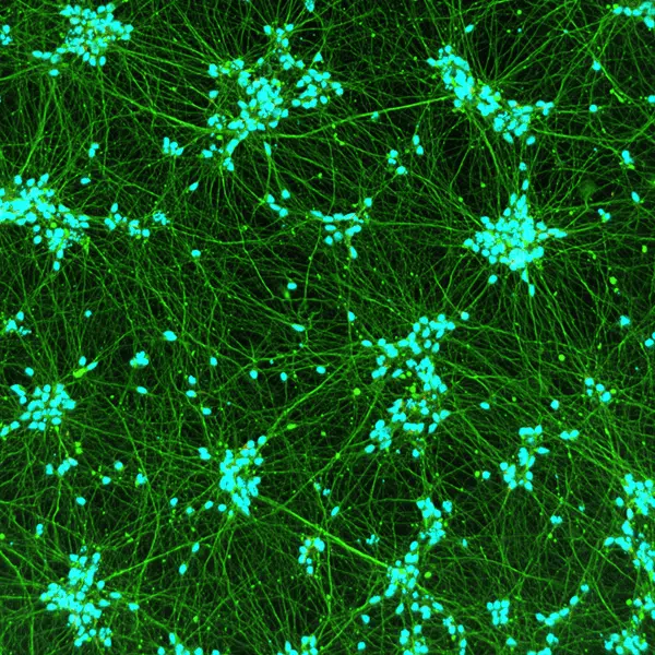
iPSC-derived GABAergic neurons express neuronal markers and display typical neurite growth. Immunofluorescent staining of Quick-Neuron™ GABAergic – mRNA Kit cell cultures shows typical neurite growth and expression of the pan-neuronal marker TUBB3 and the GABAergic neuron-specific marker PVALB on day 10 post-differentiation. Staining conditions: Anti-β-III tubulin monoclonal antibody (R&D Systems, Catalog Number: MAB1195, 1:250 dilution) in combination with a secondary antibody (Invitrogen,Catalog Number: A32723 Goat anti-Mouse IgG (H+L) Highly Cross-Adsorbed Secondary Antibody, AlexaFluor Plus 488, 1:500 dilution). Anti-PVALB primary antibody (Novus Biologicals, Catalog Number: NB120-11427 , 1:1000 dilution) in combination with a secondary antibody (Invitrogen, Catalog Number: A11037 Goat anti-Rabbit IgG (H+L) Highly Cross-Adsorbed Secondary Antibody, Alexa Fluor 594, 1:500 dilution). Nuclei were counterstained with Hoechst 33342.

iPSC-derived GABAergic neurons express neuronal markers and display typical neurite growth. Immunofluorescent staining of Quick-Neuron™ GABAergic – mRNA Kit cell cultures shows typical neurite growth and expression of the pan-neuronal marker TUBB3 and the GABAergic neuron-specific marker GAD65/67 on day 11 post-differentiation (scale bar = 100 μm). Staining conditions: Anti-β-III tubulin monoclonal antibody (R&D Sys- tems, Catalog Number: MAB1195, 1:250 dilution) in combination with a secondary antibody (Invitrogen,Catalog Number: A32723 Goat anti-Mouse IgG (H+L) Highly Cross-Adsorbed Secondary Antibody, AlexaFluor Plus 488, 1:500 dilution). Anti-GAD65/GAD67 primary antibody (Fisher Scientific, Catalog Number: PA5-36080, 1:400 dilution) in combination with a secondary antibody (Invitrogen, Catalog Number: A11037 Goat anti-Rabbit IgG (H+L) Highly Cross-Adsorbed Secondary Antibody, Alexa Fluor 594, 1:500 dilution). Nuclei were counterstained with Hoechst 33342.
Assessment of the GABA Expression Level of Quick-Neuron™ GABAergic Neuronal Culture

GABA expression level was determined by ELISA from Quick-Neuron™ GABAergic neuronal culture, using supernatants collected at day 14 and day 21 post differentiation (n=2). Data is expressed as the mean of 2 biological replicates ±SD. Supernatants of Quick-Neuron™ Excitatory neuronal culture at day 21 post differentiation were used as negative control.
Product Specifications
| Parameters | Specifications |
|---|---|
| Product Name | Quick-Neuron™ GABAergic - mRNA Kit |
| Catalog No. | GA-mRNA |
| Product Components | QNG-mRNA-P, Component N, Component P, Component G1, Component G2, and Coating Material A |
| Storage Conditions | mRNA should be stored at -80°C. All other components can be stored at -20°C or -80°C. |
| Cell Type | GABAergic Neurons |
| Induction Method | Transcription factors delivered by synthetic mRNA |
| Differentiation | At day 7 post-differentiation (CW50065) >80% TUBB3+, >50% GAD65/67+/TUBB3+, >50% PVALB+/TUBB3+ |
| Sterility | No growth observed |
| Mycoplasma | No DNA detected |
| Restricted Use | For Research Use Only. Not for use in diagnostic or therapeutic procedures. |
Resources
Quick-Neuron™ GABAergic – Human iPSC-Derived Neurons
Quick-Neuron™ GABAergic – mRNA Kit
Transcription Factor-Based Rapid Differentiation of Human iPSCs into Inhibitory, Excitatory and Sensory Neurons (Society for Neuroscience 2022)
Development of Electrophysiological Toxicity Assay System with iPSC-derived GABAergic Neurons Generated by a Rapid Differentiation Method (Safety Pharmacology Society 2023)
Establishment of High-Throughput Toxicity Assay System with iPSC-derived GABAergic Neurons Generated by a Rapid Differentiation Method (Safety Pharmacology Society 2023)
No results found.
Related Products

FAQs
Does Quick-Tissue™ technology leave a genetic footprint?
Sendai virus (SeV) is an RNA virus, so it does not integrate into the genomic DNA. In principle, a foreign gene introduced intracellularly in the form of RNA is quickly translated and expressed because, unlike DNA, RNA does not need to enter the nucleus for forced expression, thereby providing no chance of mutagenesis. This is discussed in the following review paper: Yamamoto, et al., (2009) “Current prospects for mRNA gene delivery.” Eur. J. Pharm Biopharm 71, 484-489.
Will SeV remain active after differentiation?
No. The SeV used in our kits is a temperature-sensitive mutant that is active at 33℃ but becomes inactive at 37℃, which is the temperature instructed in the user guides post-differentiation.
Is Sendai virus (SeV) dangerous?
SeV is not pathogenic to humans (i.e., humans are not the natural host of the virus) and the infection does not persist in immunocompetent animals. Furthermore, SeV used in our kits does not produce infectious viral particles upon transduction to host hPSCs and is a temperature-sensitive mutant, such that it is active at 33℃ but becomes inactive at 37℃. However, because SeV can be transmitted by aerosol and contact with respiratory secretions and is highly contagious, appropriate care must be taken to prevent potential mucosal exposure to the virus. Our SeV-based kits must be used under Biosafety Level 2 (BL-2) containment with a biological safety cabinet or a laminar flow hood and with appropriate personal protective equipment. In the event that the virus comes into contact with skin or eyes, decontaminate the affected area by flushing with plenty of water and follow the safety manual prepared by your laboratory and approved by your Institutional Biosafety Committee.
Do I need a license agreement for any of Ricoh Biosciences’ products?
No. You don’t need any licence or material transfer agreement (MTA) to use our differentiation kits or iPSC-derived cells. However, please be advised that these products are for research use only.
No results found.
Contact Us
Have a question about our products, services, or custom projects? Our team is here to help—reach out and we’ll get back to you as soon as possible.
Subscribe
Sign up to our eNewsletter to stay up to date with the latest product launches, promotions, and receive expert tips.
By signing up you are agreeing to our Privacy Policy